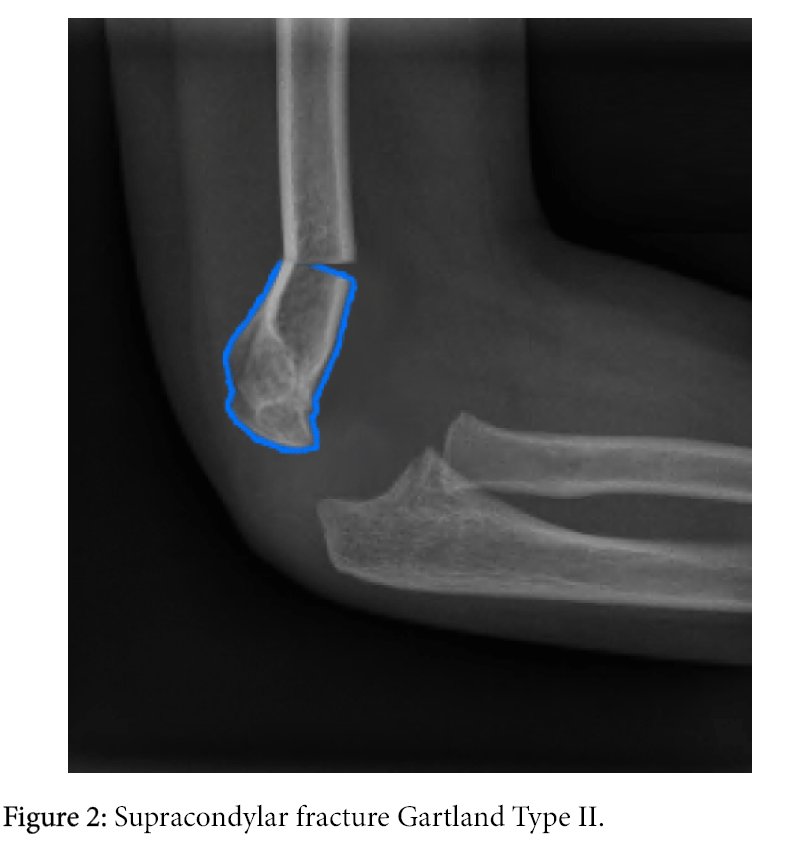

There are several key elements to address when evaluating a plain radiograph for supracondylar fractures, as not all are readily evident. Therefore, when a supracondylar fracture is present, one should have a low threshold to image other sites of clinical concern, such as the wrist or forearm, when clinically indicated by physical examination, or when physical examination is difficult, especially in young children. Associated fractures of the wrist and forearm are most common, but fractures of the proximal humerus or clavicle also occur. The clinician should bear in mind the possibility of additional associated fractures with a supracondylar fracture. , Oblique views are not typically required but can be used to further identify minimally displaced fractures if there is strong clinical suspicion for a fracture. , The lateral view is obtained with the elbow in flexion at a true 90-degree angle, or at least as close to this as possible. Proper evaluation of suspected supracondylar fractures requires anterior-posterior and lateral views of the elbow. He is grossly neurovascularly intact and there is no other apparent injury. His physical examination is remarkable for impressive right elbow swelling and pain.

He complains of right elbow pain but denies neck or back pain. The fall was witnessed by several teachers and friends, who reported no loss of consciousness or other injuries. A 5-year-old male is brought for right elbow pain and swelling after falling while playing on the monkey bars at school.


 0 kommentar(er)
